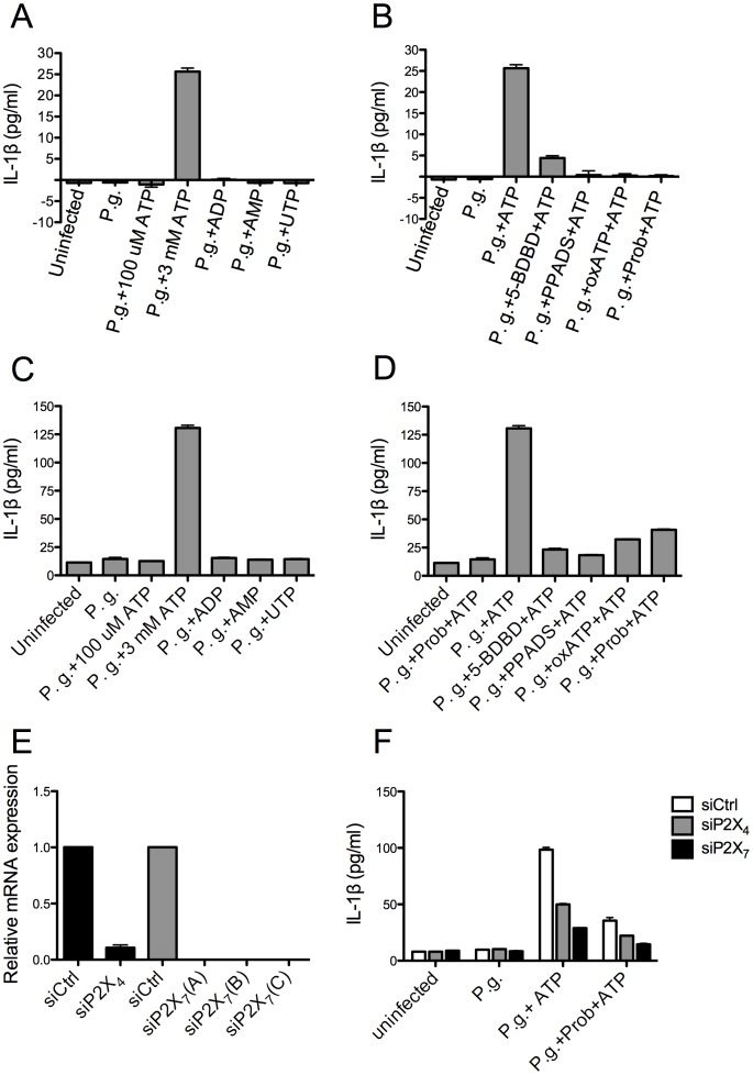
Figure 6. Abrogation of ATP-induced IL-1β secretion in P. gingivalis-infected GEC by inhibition of P2X4, P2X7, or pannexin-1.
Primary GEC (C and D) and immortalized GEC (A and B) were infected with or without P. gingivalis (P.g.) at an M.O.I. of 100 for 6 hours, followed by treatment with different pharmaceutical agents. Infected cells were treated with 100 µM ATP, 3 mM ATP, 3 mM ADP, 3 mM AMP, or 3 mM UTP individually for 1 hour (A and C). Alternatively, infected cells were pre-treated with 50 µM 5-BDBD for 15 minutes, 100 µM PPADS for 15 minutes, 100 µM oxATP for 30 minutes, or 1 mM probenecid for 10 minutes, followed by treatment with 3 mM ATP for 1 hour (B and D). The supernatants were collected and subjected to ELISA to measure IL-1β secretion. (E) Primary GEC were transfected with siRNA sequences against P2X4 or P2X7 for one day, and mRNA levels were detected by qPCR. (F) Primary GEC depleted of P2X4 or P2X7 were infected with P. gingivalis (P.g.) and treated with probenecid and 3 mM ATP as shown in (B). IL-1β secretion in the supernatants was analyzed by ELISA. The values showed averages and SD from duplicate samples, which were obtained from three separate experiments.

