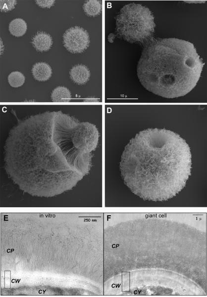
Figure 2. Morphological features of titan cells shown by electron microscopy.
A-D) Scanning electron microscopy of typical cells grown in Sabouraud medium (A) or of titan cells isolated from the lungs of infected mice (B-D). Scale bar in B applies to panels C and D. E-F) Transmission electron microscopy of cells grown in vitro (E) or of titan cells (F). CP, capsule; CW, cell wall; CY, cytoplasm. The pictures shown in this figure are reproduced from reference (16).

