Fig. 2.
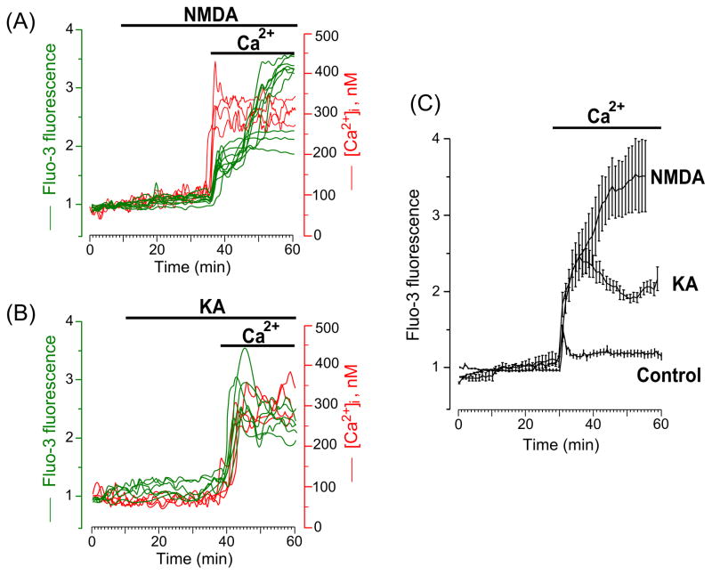
Intracellular Ca2+ signals induced by GluR agonists occur only when Ca2+ is present in the external solution. (A) Time course of the [Ca2+]i response after application of 30 μM NMDA in Ca2+-free extracellular solution, followed by the addition of 2 mM Ca2+ to the extracellular solution in the continued presence of 30 μM NMDA (the application episodes are marked with lines above the traces). Neurons were loaded with Fluo-3 (left ordinate, relative fluorescence intensity, green lines) and Fura-2 (right ordinate, [Ca2+]i, red lines). Each trace represents the response of one neuron. Data from two experiments (one with Fluo-3 and one with Fura-2) are plotted. Four (N=4) experiments for each Ca2+ indicator were performed. (B) Time course of the [Ca2+]i response after application of 30 μM KA in Ca2+-free extracellular solution, followed by the addition of 2 mM Ca2+ to the extracellular solution in the continued presence of 30 μM KA (the application episodes are marked with lines above the traces). Neurons were loaded with Fluo-3 (left ordinate, relative fluorescence intensity, green lines) and Fura-2 (right ordinate, [Ca2+]i, red lines). Each trace represents the response of one neuron. Data from two experiments (one with Fluo-3 and one with Fura-2) are plotted. Four (N=4) experiments for each Ca2+ indicator were performed. (C) Comparison of average time courses of the [Ca2+]i responses obtained in experiments with NMDA (N = 3, total number of analyzed neurons is 40), KA (N = 4, total number of analyzed neurons is 48) and in the absence of agonists (control, N = 4, total number of analyzed neurons is 83) upon an addition of 2 mM Ca2+ to the Ca2+-free extracellular solution. Mean values ± s.e. are plotted.

