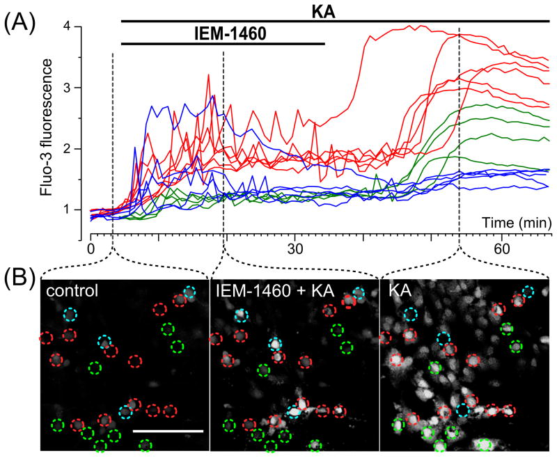
Fig. 4.
Application of KA and IEM-1460, a subunit specific open channel blocker of AMPARs reveals three different types of intracellular Ca2+ responses in cultured cortical neurons. (A) Ca2+ responses upon application of 30 μM KA with 3 μM IEM-1460 followed by IEM-1460 washout (the application episodes are marked with lines above the traces). Neurons were loaded with Fluo-3 (ordinate, relative fluorescence intensity). Each trace represents the response of one neuron. Three (N = 3) experiments were performed. (B) Fluorescent images taken at different stages (indicated by dashed lines) of the experiment illustrated in (A). Ca2+ responses of neurons marked by circles are plotted in panel A. Red, blue and green traces (in A) were obtained from neurons marked by circles of the same color. Scale bar is 100 μm and valid for all images. Detailed description and interpretation of these data are presented in sections 3.4. and 4.4.

