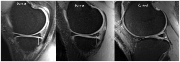
Figure 2.

Sagittal 7T MR image (TR/TE = 3270 ms/26 ms, 0.357 mm × 0.357 mm × 3 mm) demonstrating Stoller grade 3 (left panel) and grade 2 (middle panel) signal within the posterior horn of the medal meniscus in asymptomatic dancers. The meniscus from a healthy control is shown for comparison (right panel).
