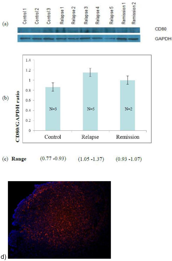
Figure 5.
Cellular podocyte extracts were probed with antiCD80 antibody to quantify CD80 protein level after podocyte culture stimulation with sera from 5 MCD patients in relapse, 2 MCD in remission, and 3 normal controls. Western blot bands from patients and normal controls a) were quantified by densitometry and results normalized using GADPH (glyceraldehyde 3 phosphate dehydrogenase) (p<0.05 between relapse and normal controls) b). Densitometry CD80 result range is given in c). CD80 in human tonsil: CD80 is expressed (red stain) in normal human tonsil using the same CD80 goat antibody (R&D Systems) as in western blots. CD80 and anti-CD80 complexes were visualized as previously described [7] d).

