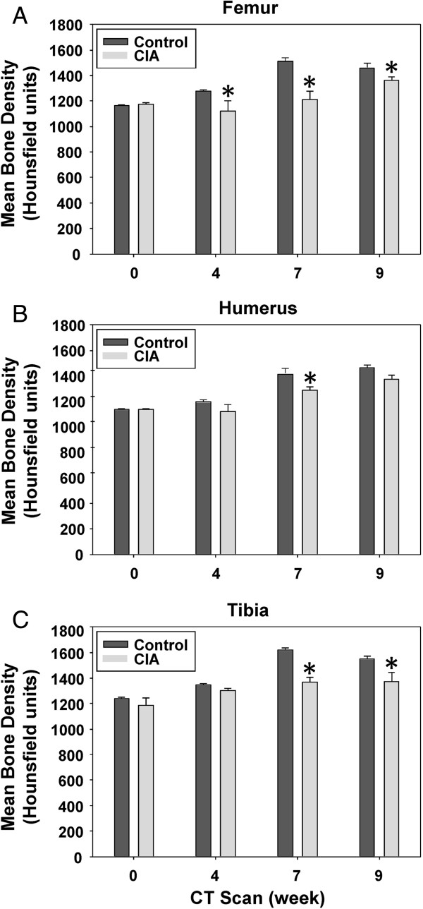
Figure 3.
Mean bone density in the bones of control and CIA mice. Shown are the mean densities and standard errors over time for (A) femur, (B) humerus, and (C) tibia from control and CIA mouse groups. P values were calculated by comparing each pair of control and CIA densities for each scan time (week 0, week 4, week 7, and week 9), and statistical significance was considered significant (*) if P < 0.05.

