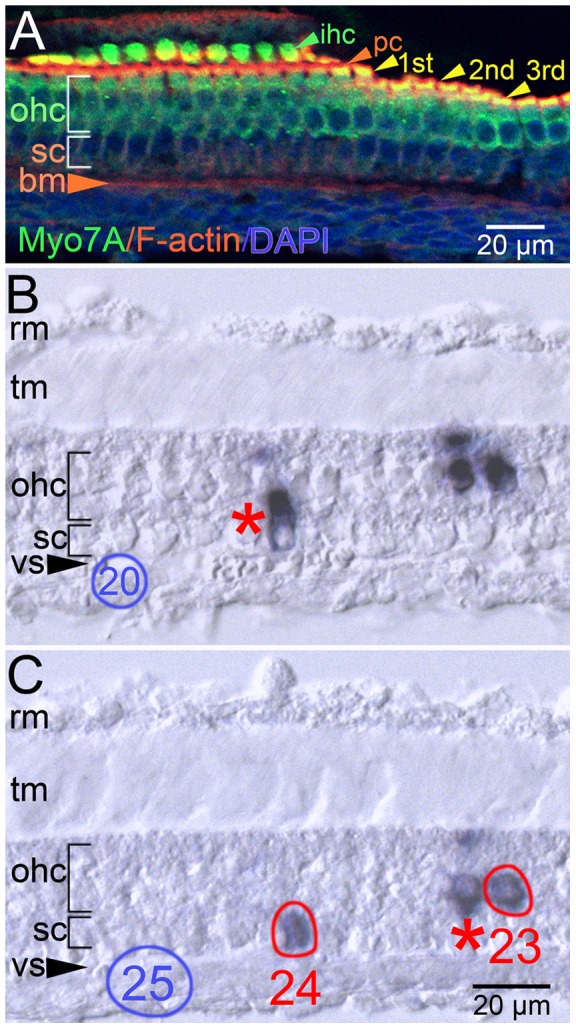
Figure 4. Outer hair cells and a supporting cell type are clonally related.

A, confocal projection of an oblique, 14 µm section of a control P6 cochlear duct. Sensory hair cells (myosin7a [Myo7A], green), filamentous actin (F-actin, red), and cell nuclei (DAPI, blue) are detectable in this atypical histological presentation of the organ of Corti. The Myo7A-positive inner hair cells (ihc) are separated from the 1st, 2nd, and 3rd rows of Myo7a-positive outer hair cells (ohc, wide bracket) by the F-actin-enriched apices of pillar cells (pc). The supporting cell (sc) nuclei occupy a region beneath the Myo7a-positive outer hair cells (ohc) indicated by the narrow bracket. B, C, oblique,14 µm, serial cryostat sections of a P6 organ of Corti in a similar orientation as in panel A presenting PLAP-positive cells of a clone. The supporting cell in pick 24 and the outer hair cell in pick 23 returned the identical sequence tag. The supporting cell in panel B (red asterisk) and the hair cell in panel C (red asterisk) did not return an oligonucleotide sequence. Negative control picks 20 (blue oval, panel B) and 25 (blue oval, panel C) did not generate a PCR product. The atypical plane of section precludes clear identification of the supporting cell type in the ohc clone. Abbreviations: bm, basilar membrane; rm, Reissner's membrane; tm, tectorial membrane; vs, vas spirale (tangential section).
