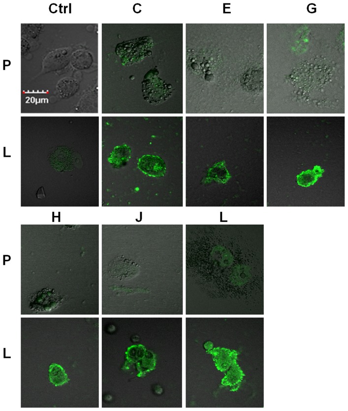
Figure 3. Immunofluorescence microscopy of phage clones from round 3 of panning.
Randomly selected PSMA-targeted phage clones (C, E, G, H, J, L) from the third round of screening bound specifically to LNCaP cells (“L”), but not PC3 cells (“P”). While staining was preferentially bound at cell surface, there was evidence of internalization (scale 20 µm).

