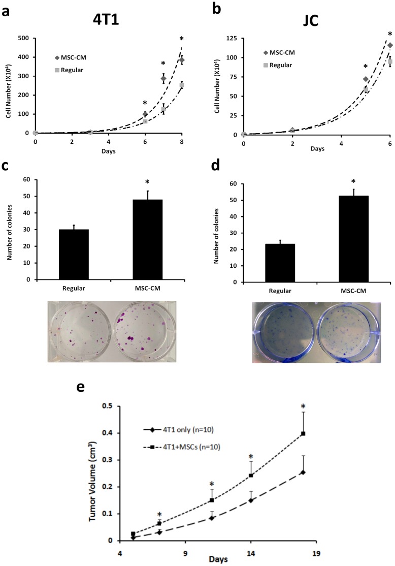
Figure 1. MSC enhances 4T1 cell proliferation and tumor growth.
(a,b) To assess cell proliferation, 4T1 or JC cells were cultured in RPMI-1640 medium or MSC-CM with the medium changed every 2 days. Cells were counted from 6 wells at each time point after culture at the indicated time points. Cells cultured in CM proliferated more than those cultured in regular medium. (c, d) To carry out the colony forming assay, 500 4T1 or JC cells were seeded and cultured using RPMI-1640 medium or MSC-CM. The colonies were stained with crystal violet after being cultured for 6 days. The cells grown using MSC-CM showed greater potential to form colonies. (c) 5×10⁵ GFP-expressing 4T1 cells alone or premixed with an equal amount of RFP-expressing MSCs were injected subcutaneously into the mammary fat pad of nude mice. Each group consisted of ten mice. Tumor growth was measured twice a week and the data are shown as the mean ± SD. Tumors derived from a mixture of 4T1 cells and MSCs had significantly enhanced growth compared to tumors derived from 4T1 cells alone. All data are shown as the mean ± SD. “*” represents a significant difference of p<0.05.

