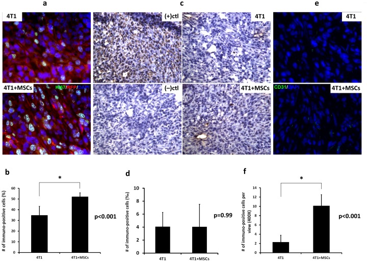
Figure 2. Tumor immunostaining for proliferation, apoptosis and angiogenesis.
5×10⁵ GFP-expressing 4T1 cells alone or premixed with an equal amount of RFP-expressing MSCs were injected subcutaneously into the mammary fat pad of nude mice. Tumors were excised for subsequent sectioning and immunostaining. Tumor sections from a 4T1 tumor or a 4T1+MSCs tumor were stained with antibody raised against Ki67 (a), CD31 (e) or subjected to TUNEL assay (c). (b, d and f) Quantification of Ki67, CD31 and TUNEL staining of tumor sections from a 4T1 tumor or a 4T1+MSCs tumor. Data represent mean values ±SD (n = 3). “*” represents a significant difference of p<0.001.

