Figure 3.

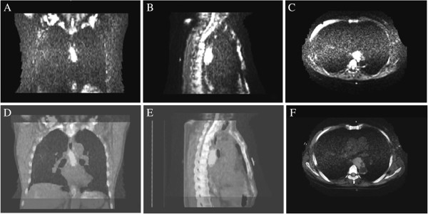
DWI scans and fused images. A, B and C show the coronal, sagittal and transverse images of DWI scans; D, E and F show the coronal, sagittal and transverse images of fused images.

DWI scans and fused images. A, B and C show the coronal, sagittal and transverse images of DWI scans; D, E and F show the coronal, sagittal and transverse images of fused images.