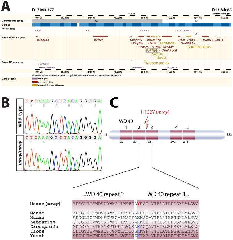
Figure 3. Positional mapping and identification of the causative mutation in the Pak1ip1 gene.
A, Ensembl-generated (version 67.37, NCBIm37) image of the genomic interval between markers D13Mit177 and D13Mit 63 the manta-ray (mray) mutation was mapped to. The coding sequences of all Ensembl/Havana genes shown in this diagram were sequenced but only one mutation was found, in the Pak1ip1 gene (boxed). The sequenced genes list from proximal to distal as follows: Slc35b3, Ofcc1, Gm9979, Tfap2a, Gcnt2, A730081D07Rik, Pak1ip1, Tmem14c, Mak, Gcm2, Sycp2l, Elovl2, BC024659, GM17364, Nedd9, Tmem170b, Gm5082, 9530008L14Rik, Hivep1, Edn1. Angled brackets indicate gene directionality. B, Sequence chromatograms of wild-type and mray PCR amplicons showing the position of the mutation (C to T) in the trace. C, Schematic structure of the Pak1ip1 protein with WD 40 repeats indicated as red boxes and the first residue of every WD 40 repeat indicated below. The mutation leads to the substitution of a histidine residue by a tyrosine residue in the first position of the third WD 40 repeat. This position of the protein is entirely conserved as shown by the sequence alignment of six species below.

