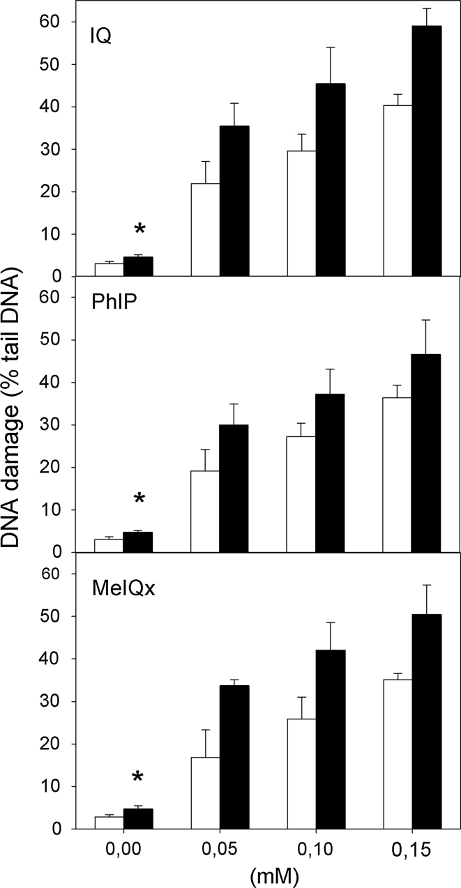
Fig. 2.
DNA damage measured as the percentage of DNA in the tail of a comet in the alkaline comet assay of H. pylori-infected (black column) and non-infected (white column) gastric mucosa cells exposed for 1 h at 37 °C to 2-amino-3-methylimidazo[4,5-f]quinoline (IQ), 2-amino-1-methyl-6-phenylimidazo[4,5-b]pyridine (PhIP) and 2-amino-3,8-dimethyl-imidazo 4,5-f]quinoxaline (MeIQx). 17 individuals were analyzed in the infected group and 23 in the non-infected (control) group. The number of cells scored for each individual was 50 and the analysis was repeated three times. Error bars denote SEM,*p < 0,001

