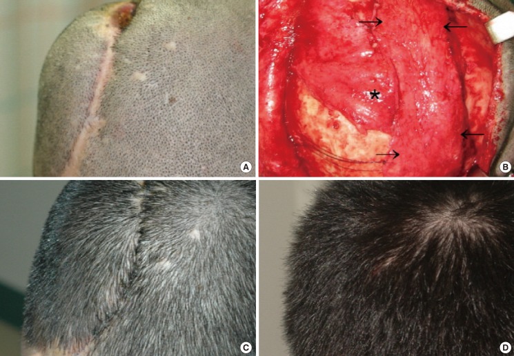
Fig. 3.
Case 3
(A) A chronic ulcer and scalp contraction along the incision line. 4 months after cranioplasty. (B) A bipedicled pericranial flap (black arrows) and an additional unipedicled flap (*) were transposed to the defect area. (C) Postoperative view at 11 days with a good scalp contour. (D) Postoperative view at 4 months.

