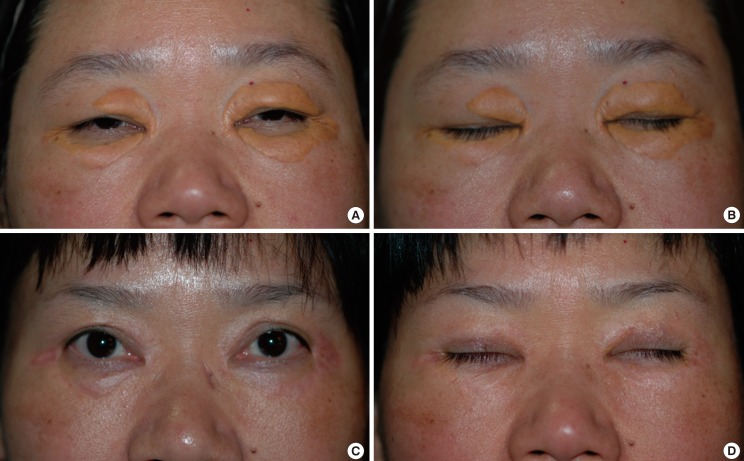
Fig. 3.
Grade IV diffuse lesion
(A, B) Preoperative photo of a grade IV diffuse lesion involving the medial and lateral sides of both upper and lower eyelids. (C, D) Postoperative photo at 1 year after surgical excision by upper blepharoplasty, lower lid orbicularis myocutaneous advancement flap, and full-thickness skin graft with excised skin from blepharoplasty. Postoperative photos show complete excision of the lesions with relatively minimal scarring and no sign of recurrence. The patient expressed satisfaction with the overall aesthetic result.

