Figure 4.
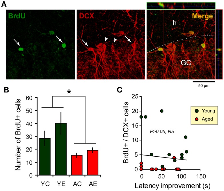
Newborn cells in the dentate gyrus (DG) over the period of the spatial memory task as examined through bromodeoxyuridine (BrdU, green) and doublecortin (DCX, red) immunostaining. (A) Newborn cells (arrows) in the DG co-express DCX (arrowheads), a marker of immature neurons. Flaps on the top and at the left of the main photograph show images merged from orthogonal planes that run through the red and green lines of the main photograph. (B) Quantification of BrdU-positive cells in the hippocampus shows an increase in the number of new cells added over a similar period of time for young-epileptic (YE) group compared to young controls (YC) and aged animals (AC, aged control; AE, aged epileptic). *P < 0.05 comparing young to aged groups (ANOVA, followed by SNK post hoc test). Values are expressed as the mean ± SEM. (C) No correlation was found between the number of BrdU+/DCX+ cells and the performance 7 days after training in the water maze (latency improvement). Data from young and aged animals are represented in different colors for clarity purposes (correlation curve not shown,P > 0.05).

