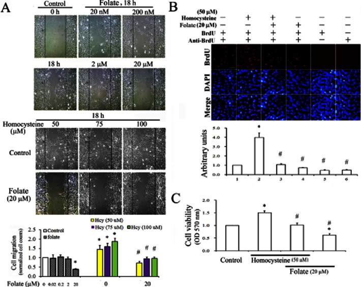
Figure 1.

Anti-proliferative and anti-migratory effects of folate on homocysteine-treated RASMC. (A) Cells cultured in IBIDI culture inserts were treated with 2 nM–20 μM of folate alone to determine optimal concentrations for its anti-migratory effect in RASMC. Thus, cells were challenged with 50–100 μM homocysteine for 30 min, followed by 20 μM folate treatment for 18 h. Cell migration was observed using a microscope with a CCD camera attached. Magnification 100×. (B) Following various treatments, DNA synthesis in RASMC was detected using a BrdU incorporation assay. The BrdU-labelled nuclei are shown as bright red spots, and DAPI staining in the same fields revealed the positions of cell nuclei. We recorded micrographs of representative fields. Magnification 400×. Results in (A) and (B) are representative of three independent experiments. (C) Cells plated in 24-well plates (5 × 10⁵ cells per well) or cultured on coverslips were challenged with 50 μM homocysteine for 30 min, then incubated with 20 μM folate for 24 h. The viability of cells was determined using an MTT assay. Data shown are the mean ± SD of five independent experiments. *P < 0.05 vs. the control; #P < 0.05 vs. the homocysteine-challenged group.
