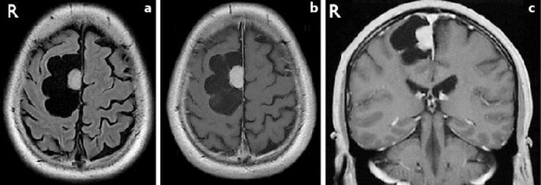
Fig. 1.
a Fluid-attenuated inversion recovery (FLAIR) image shows a cyst within the right frontal lobe. The cystic component shows an intensity that is similar to cerebral spinal fluid. A small round mass can be seen, which appeared to be a mural nodule located at the cerebral falx. Perifocal edema was not evident. b Axial T1-weighted gadolinium-enhanced image shows that the tumor is enhanced homogeneously together with its attached dura mater, but the cyst wall is not enhanced. c Coronal T1-weighted gadolinium-enhanced image.

