Figure 3.
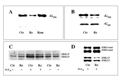
Western analysis of the effects of ribozyme on Gi2α and ERK1/2. Gi2α ribozyme (Rz) or non-cleaving Gi2α ribozyme (Rzm) complexed with DOTAP giving final ribozyme concentrations of 2.5 μM, or only DOTAP (Ctr) were added to hepatocyte cultures at 4–5 hours after the time of seeding. A: Expression of Gi2α protein was assessed after 45 h of ribozyme treatment using antibody (from Calbiochem) directed against C-terminal end of Gi1/i2α . B: Expression of Gi2α and Gqα protein levels in the same samples subsequent to 30 h of ribozyme treatment using antibodies (from NEN™ Life Science Products) against C-terminal sequences of Gi1/i2α or Gqα, respectively. The polyclonal antibodies used to assess Gi2α recognize both the α subunit of Gi2 and Gi1. As shown previously hepatocytes do not express Gi1α [19], so the reactivity with these antibodies reflects only the Gi2α levels. C, D: After 45 h of ribozyme treatment cells were stimulated with or without PGF2α (10 μM) for 5 min before they were harvested. Immunoblot using antibody against dually phosphorylated ERK1/2 (i.e. ERK1/2-P) (C) is depicted. In Fig. D is developed images from the same immunoblot using antibody detecting total amount of ERK1/2 (i.e. both phosphorylated and unphosphorylated forms) (upper panel) and antibody against dually phosphorylated fractions of ERK1/2 (lower panel).

