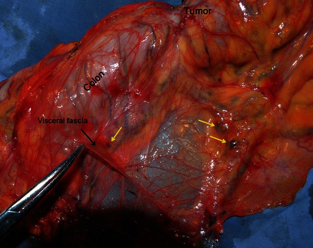
Fig. 10.

Resected specimen of ascending colon cancer after CME operation (magnification of Fig. 9B). The intact visceral fascia is in the dorsal view. The lymph nodes and supply vasculature are covered by the visceral fascia. The yellow arrows indicate black lymph nodes.
