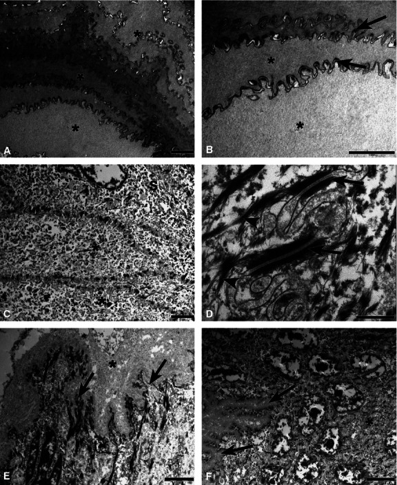
Fig. 5.

Transmission electron photomicrographs of the dorsal epithelium of the tongue of agouti. (A) In the keratinised layer, epithelial cells (*) were superimposed and arranged in parallel and connected by numerous desmosomes. (B) Epithelial cells (*) and the microridges present on the cell surface (arrows) at a higher magnification. (C) Flattened cells (*) were noted in the granular layer. (D) The desmosomes (arrows) and intermediate filaments (arrowheads) were observed at a higher magnification in the granular layer. (E) Interface region between the lamina propria and basal layer of the epithelium showing the dense collagen layer of the connective papillae (*) and their cytoplasmic projections (arrows). (F) The interdigitation (arrows) between the basal and spinous layers (*) at higher magnification. Scale bars: 2 μm (A,-C,E), 0.5 μm (D) and 5 μm (F).
