
Fig. 2.
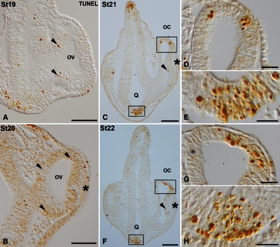
TUNEL-positive bodies in the developing visual system of small-spotted catshark embryos during St19 (A), St20 (B), St21 (C-E) and St22 (F-H). Frontal cryosections were treated according to the TUNEL method. Lens placode is indicated in panels (B,C,F) by asterisks. Boxed areas in (C) and (F) are shown at higher magnification in (D,E) and (G,H), respectively. In all panels, dorsal is up. During these early stages, sparse TUNEL-positive bodies were observed in the neuroepithelium of the optic anlage (arrowheads in A,B) and in the neuroepithelial cells of the presumptive neural retina (arrowheads in C,F). Areas with abundant TUNEL-positive bodies were detected by St21–22 in the dorsal-most part of the optic cup (C,D,F,G) and in the presumptive optic chiasm (C,E,F,H). oc, optic cup; ov, optic vesicle; Q, optic chiasm. Scale bars: 200 μm (A-C,F); 50 μm (D,E,G,H).