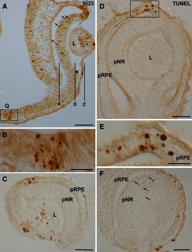
Fig. 3.

Spatial distribution of TUNEL-positive bodies in the developing visual system of small-spotted catshark embryos at St23. Frontal (A,B) and sagittal (C–F) cryosections were treated according to the TUNEL technique. (C,D,F) Overviews of sections at the level indicated in (A). Boxed areas in (A) and (D) are shown at higher magnification in (B) and (E), respectively. In all panels, dorsal is up. TUNEL-positive bodies were concentrated at the dorsal-most part of the optic cup (A,C-E), at the presumptive optic chiasm (A,B), and at the anterior region of the lens vesicle (A,C). Many TUNEL-positive bodies were dispersed throughout the undifferentiated neural retina (arrows in A,F). Notice that an undifferentiated spherical lens is present at this stage (A,C,D). L, lens; pNR, presumptive neural retina; pRPE, presumptive retinal pigment epithelium; Q, optic chiasm. Scale bars: 100 μm (A,C,D,F); 50 μm (B); 25 μm (E).
