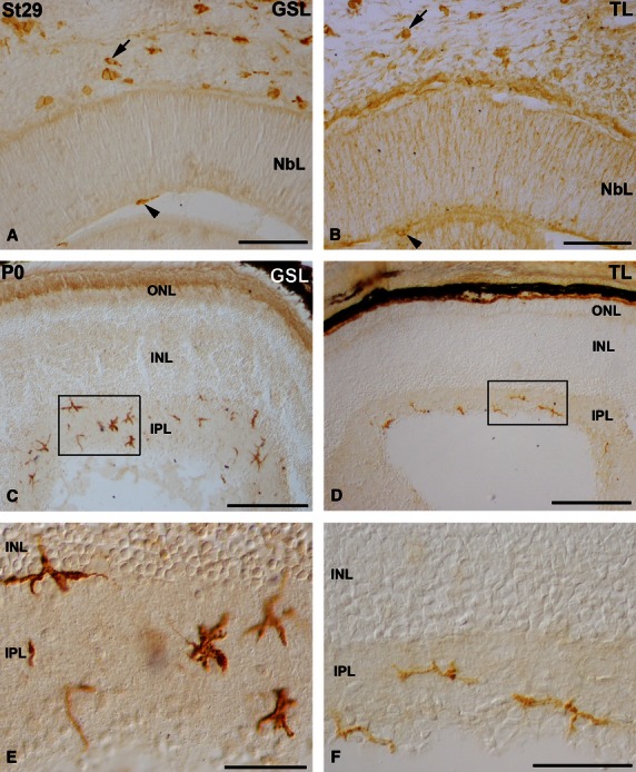
Fig. 6.

Lectin-positive cells in the retina of St29 embryos (A,B) and hatchlings (C-F) of small-spotted catshark. Cryosections were treated according to GSL (A,C,E) or TL (B,D,F) histochemistry technique. Boxed areas in (C) and (D) are shown at higher magnification in (E) and (F), respectively. Many round/amoeboid lectin-positive cells appeared in the mesenchyme (arrows in A,B) and in the vitreous cavity (arrowheads in A,B) during embryonic stages. Abundant ramified stellate lectin-positive cells were observed mainly in the IPL of the shark retina at the hatching stage (C-F). GSL, Griffonia simplicifolia lectin; INL, inner nuclear layer; IPL, inner plexiform layer; NbL, neuroblastic layer; ONL, outer nuclear layer; TL, tomato lectin. Scale bars: 100 μm (A,B); 200 μm (C,E) 50 μm (D,F).
