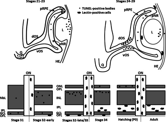
Fig. 10.

Schematic drawings showing the spatiotemporal distribution of TUNEL-positive bodies and lectin-positive cells in the developing visual system of the small-spotted catshark. During early stages, TUNEL-positive bodies are found to be accumulated in four precise and spatiotemporally distinct locations: the presumptive optic chiasm (St21–23), the dorsal-most part of the optic cup (St21–23), the lens tissue (St23–26) and the distal optic stalk (St27–29). During these early stages, TUNEL-positive bodies are sparse in the undifferentiated neural retina. However, during the period of retinal histogenesis and cell differentiation (St31 onwards), a clear vitreal-to-scleral gradient of cell death occurs, affecting cells located in the three nuclear layers. By the early stages of development (St24–31), lectin-positive cells are absent from the nervous parenchyma and are only present in the vitreous cavity of the optic anlage. From St32-early onwards, many lectin-positive cells are detectable along the entire length of the optic nerve. Furthermore, lectin-positive cells are seen to colonize progressively more scleral retinal layers as development advances, to gain access to the IPL. dOS, dorsal optic stalk; GCL, ganglion cell layer; HE, head ectoderm; INL, inner nuclear layer; IPL, inner plexiform layer; L, lens; NbL, neuroblastic layer; OFL, optic fiber layer; ON, optic nerve; ONL, outer nuclear layer; OPL, outer plexiform layer; pNR, presumptive neural retina; pRPE, presumptive retinal pigment epithelium; Q, optic chiasm; vOS, ventral optic stalk.
