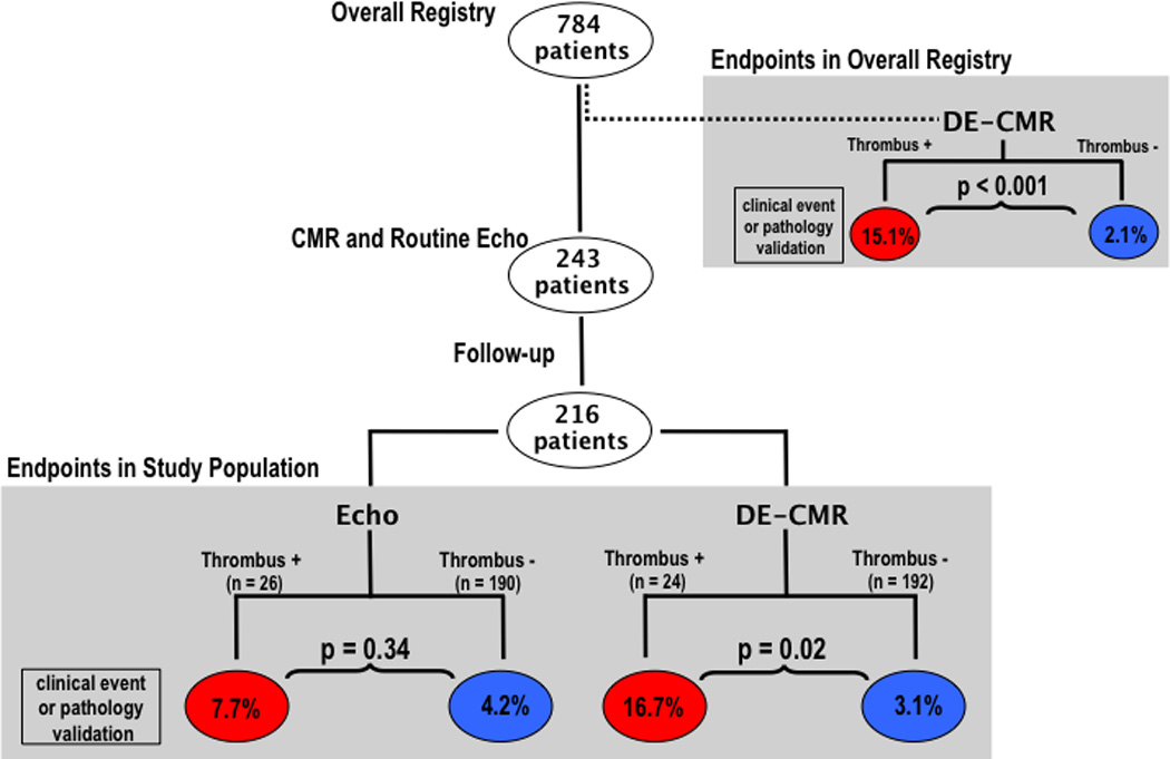
Figure 1. Follow-up Endpoints in Relation to Imaging Findings.

Stratification of patients with follow-up according to presence or absence of thrombus by DE-CMR yielded over a 5-fold difference in study endpoints (TIA, CVA, or pathology-verified thrombus) between groups whereas stratification according to echo thrombus yielded a 1.8 fold difference (red = thrombus +, blue = thrombus−).
