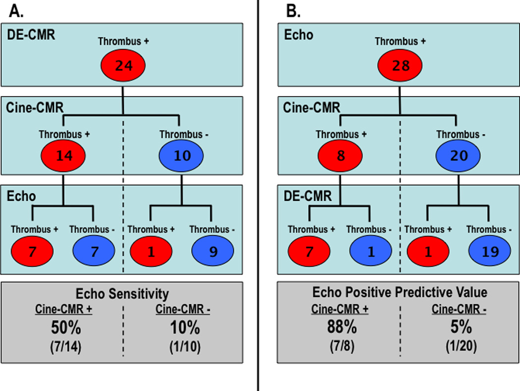
Figure 3. Imaging Results Concerning LV Thrombus.

Echo results concerning the diagnosis of thrombus stratified by cine- and DE-CMR findings (red = thrombus +, blue = thrombus −). Both echo sensitivity (3A) and positive predictive value (3B) were higher among cases in which thrombus was also evidenced by cine-CMR vs. those in which cine-CMR was negative. While cine-CMR appropriately detected thrombus in an additional 7 patients with negative echo, both tests were negative in 9/24 patients with thrombus by DE-CMR tissue characterization.
