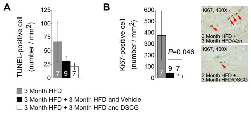
Figure 4.
Cell apoptosis and proliferation in atherosclerotic lesions from aortic arches from Ldlr−/− mice that consumed 3 months of an atherogenic diet followed by another 3 months of the same diet with and without treatment with DSCG. A. TUNEL-positive cell numbers per mm2 in aortic arch. B. Ki67-posiive cell numbers per mm2 in aortic arch atherosclerotic lesions. Representative images were shown to the right. The number of mice per group was indicated in each bar.

