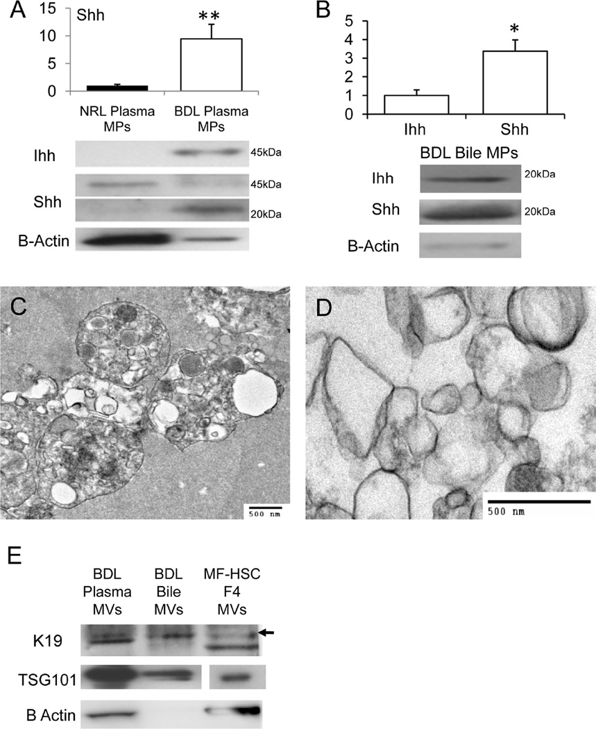
Figure 3. Bile Duct Ligation (BDL) activates release of Hh morphogens in MPs.
Western blot analysis of mature 20kDa Shh peptide in plasma MPs from sham-operated normal (NRL) and 14 days post-BDL rats (A); Western blot analysis of MPs fractions purified from bile of rats post-BDL (10ug protein/lane). Bile from NRL rats was not sufficient to isolate MPs and only bile from BDL rats was analyzed (B). Graphs demonstrate mean +/−SD results of 3 experiments. In each experiment, plasma or bile were pooled from at least 3 rats/group to obtain MPs (*P<0.05;**P<0.005); TEM images of plasma MPs (C) and biliary MPs (D) from BDL rats; Western blot demonstrating expression of K19, TSG101, and B-Actin in MPs that were isolated from the plasma and bile of BDL rats. Results are compared to expression of these proteins in an equivalent amount (10ug protein/lane) of sucrose density purified fraction 4 from PDGF-treated cultured MF-HSC (E).

