Figure 4.
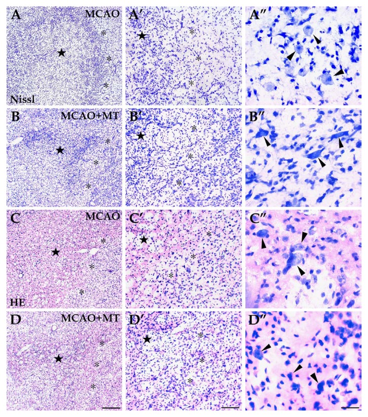
Histological changes in ischemic striatum with melatonin treatment. The infarcted area, located in the dorsolateral striatum, consisted of a dark-stained ischemic core (⋆) and a pale-stained outer zone (*) surrounding the ischemic core. Both Nissl (image sets A and B) and HE staining (image sets C and D) showed severe neuron loss and infiltration of inflammatory cells in the ischemic core whereas the neuron loss was less severe and a few large-sized neurons survived (arrowheads) in the outer zone. However, in the MCAO+MT group (image sets B and D), the ischemic core appeared smaller and more neurons survived in the outer zone than in the MCAO group (image sets A and C). Scale bars: A–D, 250 µm; A’–D’, 100 µm; A’’–D’’, 30 µm.

