Figure 6.
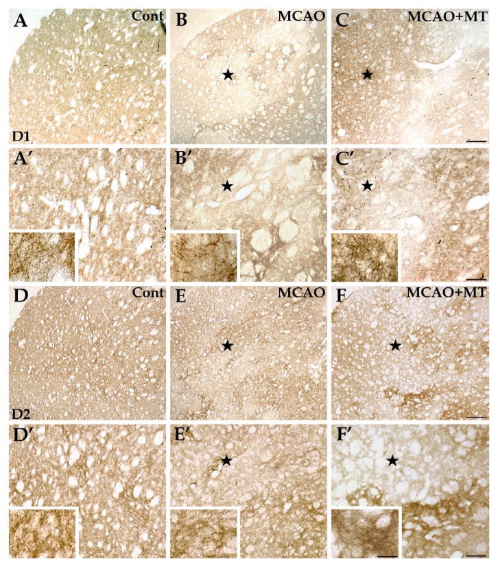
Changes of D1+ and D2+ neurons in ischemic striatum with melatonin treatment. In brain sections with immunolabeling of D1+ neurons (A, A’, B, B’, C, and C’), the immunolabeled structures, presented as neuropils, were densely and evenly distributed in the striatum of the control group (A and A’). In the MCAO group (B and B’), the D1+ neuropils were nearly absent in the ischemic core (⋆) but changes of the D1+ neurons in the outer zone of the infarct were less severe. In the MCAO+MT group (C and C’), however, the infarcted size was reduced and D1+ fibers appeared denser in the outer zone. As for immunolabeling of D2+ neurons (D, D’, E, E’, F, and F’), the immunolabeled neuropils were also densely and evenly distributed in the striatum of the control group (D and D’). The D2+ neuropils were scarce in the ischemic core (⋆) and those existing in the outer zone were aggregated as mass in the MCAO group (E and E’). In the MCAO+MT group (F and F’), the infarcted size was reduced and the density of D2+ fibers was significantly increased in the outer zone. Cont is short for control (or sham-operated). Scale bars: A–F, 250 µm; A’–F’, 100 µm; small images in A’–F’, 30 µm.

