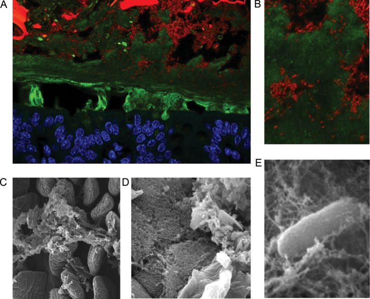
Fig. 1.
Localization of Bacteroides thetaiotaomicron (Bt) to the outer layer of colonic mucus. (A) A cross-section of distal colon of a gnotobiotic mouse colonized with Bt. Cells are stained with DAPI and false colored with blue (host tissue) or red (bacterial cells) and mucus is stained with an anti-MUC2 antibody (green). The small Bt rods occupy discrete microhabitats that include dietary plant material (intense large red and green objects), loose lumenal mucus (diffuse green signal) and the outer loose layer of epithelium-adjacent mucus. (B). Zoomed in the view of Bt embedded within mucus. Scanning electron micrographs of (C) mucus covering intestinal villi of the mouse; (D) Bt embedded in a mucus lattice (middle) overlying epithelial cells (bottom left), and in food particle (upper right), within the gnotobiotic mouse gut (Sonnenburg et al. 2005); (E) zoomed in view of Bt in mucus (Sonnenburg et al. 2005). Bt cells are ∼1 × 5 μm.

