Fig. 5.
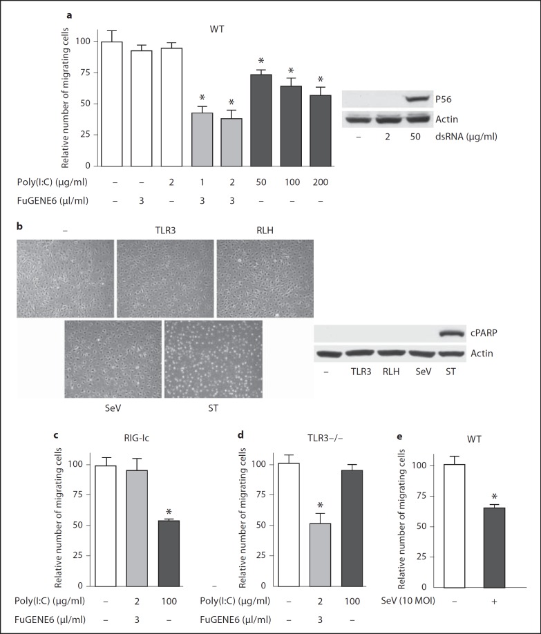
Antiviral innate immunity suppresses cell migration. a Wild-type (WT) murine podocytes were stimulated as indicated in the abscissa. Migration of cells into a defect in the monolayer in a wound-healing assay was quantified 24 h after the stimulation (left panel). Cells left untreated, incubated in FuGENE6 alone, or incubated in the presence of 2 µg/ml Poly(I:C) are in white bars; cells subject to Poly(I:C) transfection (with FuGENE6 and low-dose Poly(I:C) are in gray bars, and cells treated by adding (high-dose, extracellular) Poly(I:C) are in black bars. Murine P56 induction in WT murine podocytes was analyzed by Western blot (right panel). Cells were treated by adding extracellular Poly(I:C) at indicated concentrations for 16 h. b Murine podocytes were stimulated with added Poly(I:C) (50 µg/ml, TLR3) or transfected Poly(I:C) (RLH), infected by SeV (10 MOI) or treated with staurosporine (1 µM, ST) for 24 h. Culture fields were photographed (left) and cell lysates were analyzed for cleaved PARP (cPARP) by Western blot (right). c Migration of murine podocytes expressing a dominant negative RIG-I (RIG-Ic) was measured by a wound-healing assay as described above in a. d Migration of podocytes isolated from TLR3−/− mice was tested as described above in a. e Migration of WT murine podocytes was tested by a wound-healing assay after 24-hour incubation in culture medium (white bar) or infection by SeV (10 MOI, black bar). * p < 0.05, versus untreated cells in culture medium.

