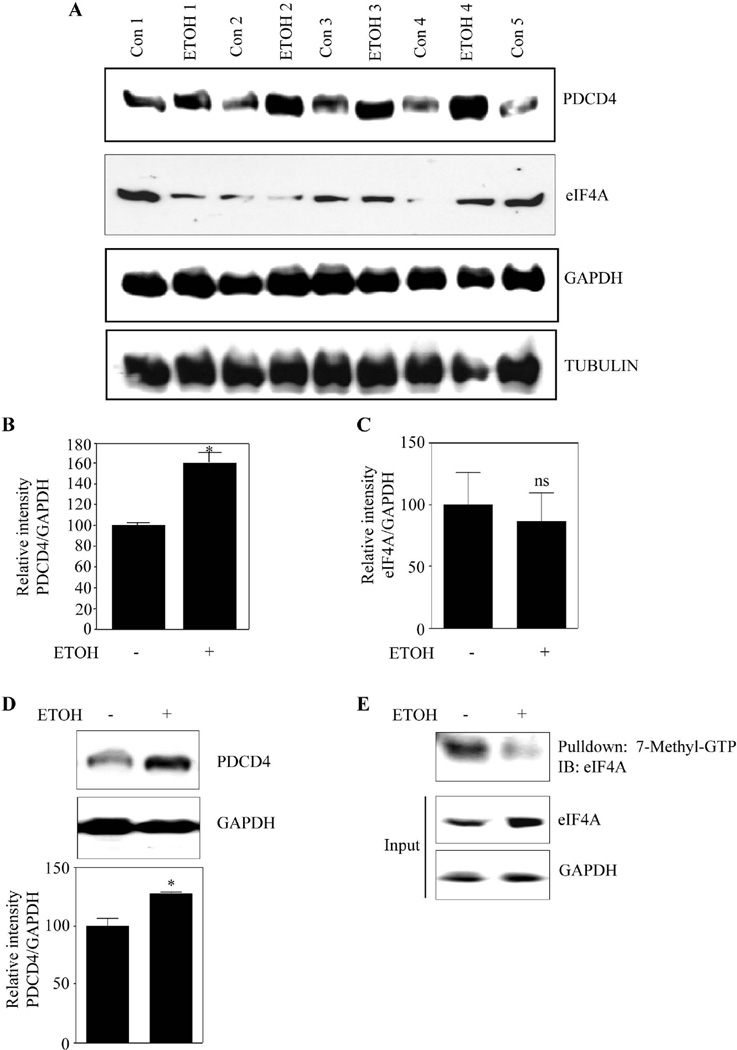
Figure 7. Prenatal ethanol exposure increases PDCD4 expression in fetal brain cortex and rest of the brain.
(A) Pregnant rats (Sprague-Dawley) at embryonic day 16 (E16) were administered ETOH (4g/kg body weight) or isocaloric dextrose by gastric intubation at 12 h intervals for two days. At E18 brain cortex from embryos were dissected and processed for PDCD4, eIF4A, GAPDH, Tubulin protein expression by immunoblotting (n=6). (B) This panel illustrates the densitometric scanning ratio of PDCD4/GAPDH intensities. Student’s t test was performed to determine the significance of treatment. * P<0.05 compared with isocaloric dextrose administered animals (mean ± s.e.m, n=6). (C) Densitometric scanning ratio of eIF4A/ GAPDH intensities. Statistical analysis was determined by Student’s t test. ns - represent not significant compared with untreated controls (mean ± s.e.m, n=3). (D) Equal amount of protein lysates from rest of the brain devoid of cortex was separated by SDS-PAGE electrophoresis and immunoblotted with PDCD4 and GAPDH. Lower panel show the densitrometric analysis of PDCD4/GAPDH band intensities. Statistical analysis using Student’s t test indicates * P<0.05 when compared with isocaloric dextrose administered animals. (E) 225µg of cortical brain lysates obtained from 7A is processed for methyl cap analysis as in Fig. 4A and a representative immunoblot is given.

