
Figure 8.
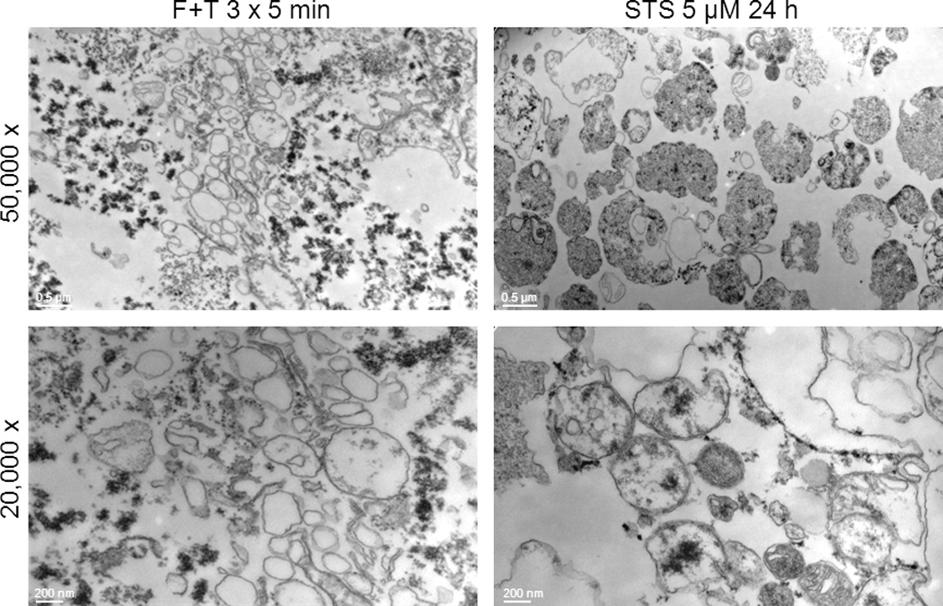
Electron microscopy of particles from necrotic and apoptotic Jurkat cells. Electron microscopy was performed as described in Material and Methods of particles obtained from either necrotic or apoptotic cells Jurkat cells subjected to either freeze-thaw or treated with staursporine at 5 µM for 24 hours. Panel A and C show particles from freeze-thaw cells while Panels B and D show particles from cells treated with staurosporine. Bars represent 200 nm and 500 nm respectively.