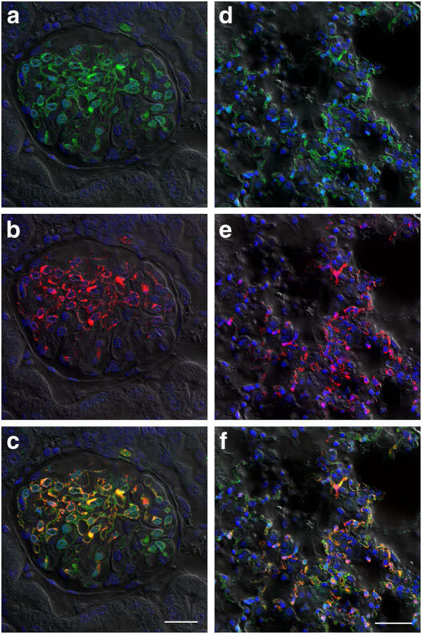
Figure 7.
Expression of GFP in tissues sectioned from HeV-GFP challenged ferrets. Ferrets were challenged with 5,000 TCID50 of HeV-GFP and succumbed to disease on days 7 and 8. At necropsy, kidney (a,b,c) and lung (d,e,f) samples were collected, fixed with paraformaldehye, sectioned and imaged by confocal microscopy. To assess sensitive of GFP expression (green; a and d) to traditional staining techniques, HeV nucleocapsid protein (red; b and e) was co-stained using rabbit anti- HeV N sera detected with anti-rabbit conjugated Alexafluor 568. Composite images (c and f) show both GFP and HeV N labelling. Scale bars: c =25 μm, f = 40 μm.

