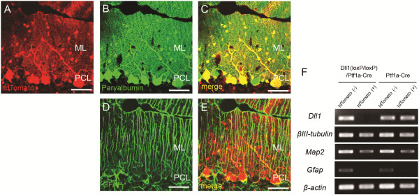
Figure 6.

Conditional ablation of Dll1 in cerebellar inhibitory neurons. (A-E) Immunohistochemical images with anti-parvalbumin (B, C green) or anti-GFAP (D, E green) antibody were overlaid with tdTomato fluorescence (A red) in Ptf1a–Cre/ROSA–tdTomato mice. (F) RT-PCR analysis shows deletion of Dll1 in tdTomato-positive inhibitory neurons from Dll1(loxP/loxP)/Ptf1a-Cre mice. Scale bars: 50 μm (A-E). ML, molecular layer; PCL, Purkinje cell layer.
