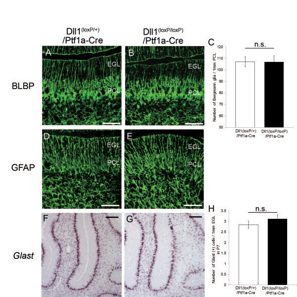
Figure 7.
The development of BG was normal in Dll1(loxP/loxP)/Ptf1a-Cre mice. (A, B) Immunohistochemistry for BLBP (green) in Dll1(loxP/+)/Ptf1a-Cre (A) and Dll1(loxP/loxP)/Ptf1a-Cre (B) mice at P7. (C) Quantification of the number of BLBP-positive cell bodies. (D, E) BG labeled by anti-GFAP (green) antibody in Dll1(loxP/+)/Ptf1a-Cre (D) and Dll1(loxP/loxP)/Ptf1a-Cre (E) mice at P7. (F, G) In situ hybridization for GLAST in Dll1(loxP/+)/Ptf1a-Cre (F) and Dll1(loxP/loxP)/Ptf1a-Cre (G) mice at P7. (H) Quantification of the number of GLAST-positive cells in EGL at P7. n.s., no significant. Scale bars: 50 μm (A, B, D, E), 250 μm (F, G). EGL, external granular layer; PCL, Purkinje cell layer.

