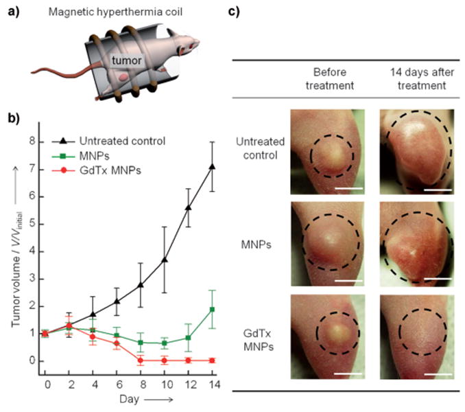
Figure 3.

In vivo magnetic hyperthermia. a) Representation of the in vivo magnetic hyperthermia study. Magnetic nanoparticles (75 μg) are directly injected into the tumor (tumor volume 100 mm3, n = 3) of a mouse and subjected to an AC field for 30 min at 43 ± 1 °C. b) Plot of tumor volume (V/Vinitial) versus the number of days after treatment. In the group treated with GdTx MNP hyperthermia (red line), the tumor is completely eliminated by day 8, whereas the MNP hyperthermia group (green line) shows only initial reduction in tumor volume, followed by regrowth. c) Images of xenografted tumors (MDA-MB-231) on nude mice before treatment (left column) and 14 days after treatment (right column). Scale bars: 5 mm.
