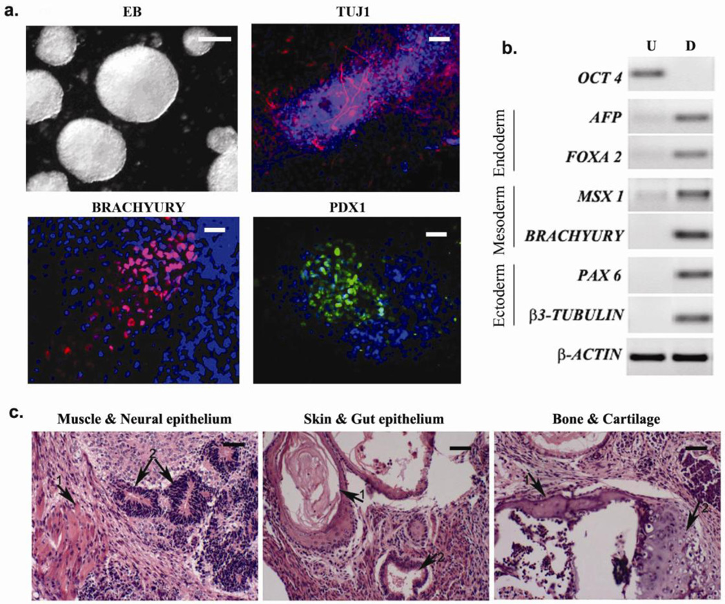
Figure 3. In vitro and in vivo differentiation of iPSCs generated with 3 compound treatment.
(a) Micrographs show embryoid bodies (EB) generated from iPSCs and in vitro differentiation into ectodermal (βIII tubulin), mesodermal (brachyury) and endodermal (PDX1) cell types. Scale bars, EB: 100 µm; others 10 µm (b) RT-PCR showing expression of representative lineage markers and the absence of OCT4 mRNA expression in differentiating cells. U- undifferentiated, D- differentiated. (c) Teratomas generated in nude mice from iPSCs (3 independent colonies tested) consist of tissues from all three germ layers. Left panel: 1- muscle, 2- neural epithelium; middle panel: 1- skin, 2- gut epithelium; right panel: 1- bone, 2- cartilage. Scale bars, 20 µm.

