Figure 1.
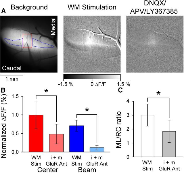
WM stimulation evokes beam-like responses. A, Left, Background fluorescence image of Crus II after injection of Oregon Green. Also shown is electrode placement within the folium and ROIs used to quantify the center (red) and beam (blue) components of the fluorescence response. Microelectrode was lowered to a depth of 500 μm below the surface. Middle, The beam-like Ca2+ response to WM stimulation (200 μA, 100 μs pulses at 100 Hz for 100 ms). Right, The beam response is largely blocked by i+mGluR antagonists (100 μm DNQX, 250 μm APV, and 200 μm LY367385). B, Summary data (mean ± SD, n = 4 mice) for the center and beam response components before and after block by GluR antagonists. *p < 0.05. C, Summary ML/RC data for WM stimulation (open bar) and after (filled gray bar) application of i+mGluR antagonists (n = 4 mice).

