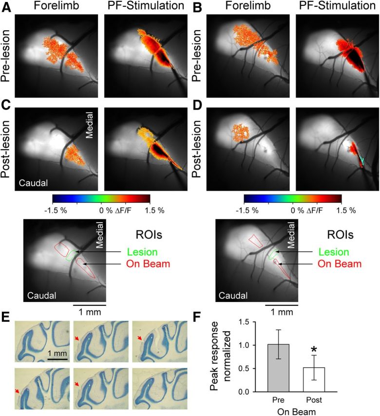
Figure 5.

PFs contribute to the beam-like response in Crus I. A, B, Example images from two mice showing the fluorescence responses evoked by forelimb stimulation (left) or direct PF stimulation (right). C, D, After electrolytic lesioning of the molecular layer (inset below, green ROIs), the peripherally evoked beam-like response (left) is reduced. Also, the response to direct PF stimulation is disrupted (right). Bottom images, The R0Is used to quantify the On Beam response (red) and the location of the lesion (green). E, Consecutive 40 μm sections showing the lesion location and depth in the molecular layer (red arrow). F, Normalized On Beam response to forelimb stimulation is significantly reduced after PF lesioning (n = 7 mice). *p < 0.05.
