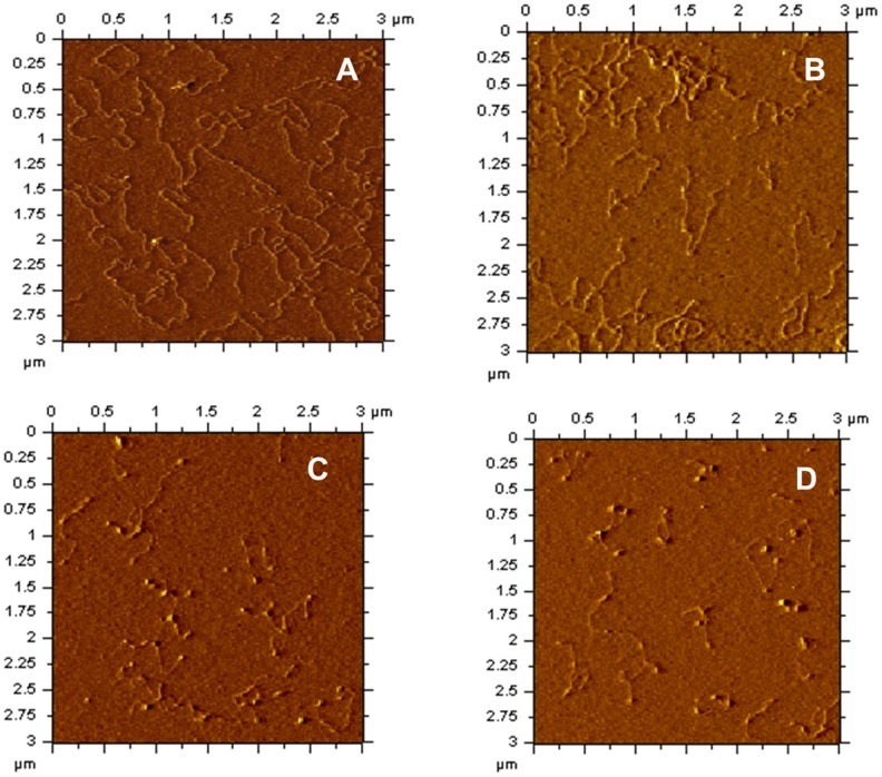
Figure 6. Representative AFM images of protein-DNA complexes formed between mIHF-80 and linear DNA.
(A) Linear pPROEX-HTc in the absence of mIHF-80. (B)–(D): mIHF-80-pPROEX-HTc (linearized) complexes with following mIHF-80 dimer molecules per 10 base pairs of DNA; (B) 2, (C) 4 and (D) 8. The scale of all images is (3 µm×3 µm).

