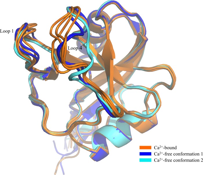
FIGURE 2.
Comparison of Ca2+-bound and Ca2+-free derCD23 structures. Superimposition of Ca2+-free derCD23 structures (colored blue and cyan) and Ca2+-bound derCD23 structures (orange) is shown. Many of the residues that undergo conformational change upon calcium binding are also involved in the interaction with IgE.

