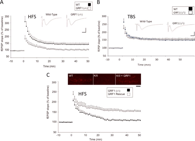
FIGURE 1.
Ras-Grf1 knock-out mice display defective HFS-induced but not TBS-induced LTP in CA1 hippocampus of 2-month-old mice. A and B, hippocampus slices from 2-month-old wild-type (filled squares) or Ras-Grf1 knock-out mice (open squares) were stimulated with HFS (A) or TBS (B). The recorded fEPSP slope using field recordings is expressed as a percentage of base line ± S.E. For HFS: WT; n = 11 slices from six mice; grf1−/−; n = 12 slices from eight grf1−/− mice). For TBS: WT; n = 8 slices from five mice; grf1−/−; n = 9 slices from five mice. Insets, representative fEPSPs recorded. C, control GFP-expressing adenovirus (filled diamonds) or GRF1-expressing adenovirus (open diamonds) was injected into the CA1 hippocampus. 9–11 days later, HFS LTP was measured in isolated hippocampal slices. Data represent the mean ± S.E. (Ras-Grf1; n = 10 slices from five mice; GFP; n = 13 slices from nine mice). Inset, CA1 hippocampus was stained with anti-RAS-GRF1 antibodies (calibration: horizontal, 10 ms, vertical, 1 mV, scale bar, 50 μm).

