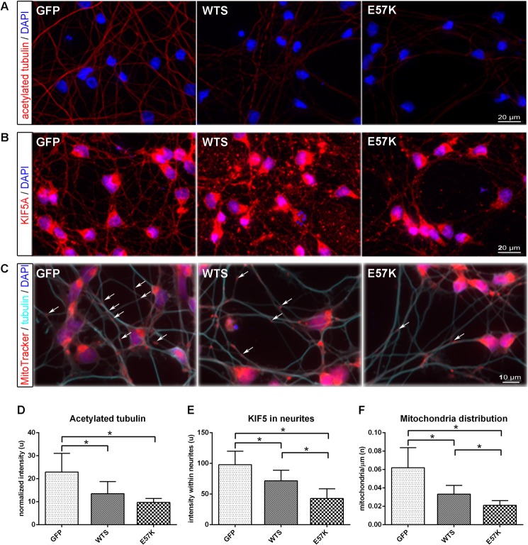
FIGURE 6.
Mild overexpression of WTS and E57K in dopaminergic human neuronal cells leads to impaired MT stability and KIF5-dependent cargo distribution. A–C, fluorescence images of differentiating LUHMES cells (day 4) infected with lentiviruses expressing either GFP or α-Syn (WTS or E57K) at MOI 2, fixed with methanol, and stained for acetylated tubulin (A), KIF5 (B), and mitochondria and β3-tubulin (C). Note that the GFP signal is not detectable anymore after methanol fixation. Scale bar, 20 μm (A and B) and 10 μm (C). D, the level of acetylated tubulin, representing a portion of stabilized MTs, is significantly reduced in WTS- and E57K-overexpressing cells. Intensity values for acetylated tubulin were normalized to the numbers of DAPI-positive nuclei. E, KIF5 signal is impaired in neurites of WTS and even stronger in E57K neuronal cells. Signals are presented as mean intensity within single traced neurite ± S.D. (error bars) At least 100 neurites were analyzed for each cell type in three independent experiments. F, significantly fewer mitochondria are present within neurites of WTS-overexpressing and much fewer in E57K-overexpressing neuronal cells. Mitochondria are visualized using MitoTracker_RedCMXRos. The number of mitochondria was determined per 1 μm of neurite length. Some mitochondria are highlighted with arrows. *, p < 0.05.

