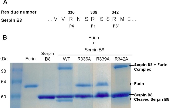
FIGURE 2.

SDS-PAGE analysis of reactions of parent and RCL variant forms of serpin B8-5S with furin. A, sequence of the reactive center loop P6–P5′ region of serpin B8-5S showing the two RXXR sequences at P4–P1 and P1–P3′. B, SDS-PAGE analysis of the reactions of parent and three single Arg → Ala variant forms of serpin B8-5S (4 μg) with furin (1 μg) as indicated. Protein bands were stained with Coomassie Blue. Labels on the right indicate the position of serpin-protease complex and cleaved serpin bands. Molecular weight standards are in the leftmost lane.
