
FIGURE 5.
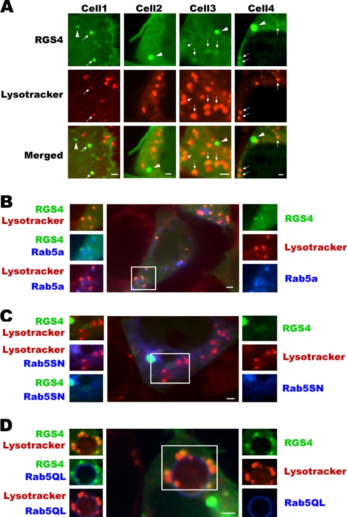
Rab5 promotes increased RGS4 co-localization with a Lysotracker® Red-labeled pool of endosomes. A, shown are four different cells transfected with RGS4-YFP and subsequently labeled with Lysotracker® Red under base-line conditions. Arrows indicate co-localized endosomes between RGS4 and Lysotracker® Red, and arrowheads indicate non-co-localized endosomes. B–D, as in the legend of Fig. 3 above, the center and surrounding images represent different channel views of cells that are marked in three colors with RGS4-YFP (green), the indicated CFP-Rab5 construct (blue), and Lysotracker® Red. For each panel, the right side images are single channel, and left side images represent overlap of two channels. All images were captured 24 h post-transfection using a spinning disc microscope with ×60 oil objective. Scale bars, 1 μm.