FIGURE 1.
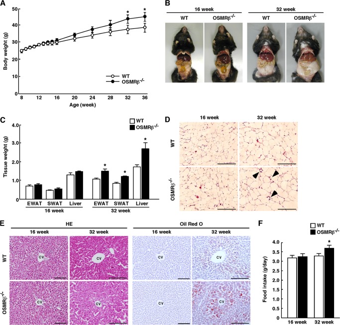
The characteristics of WT and OSMRβ−/− mice under normal diet conditions. A, shown are the body weights of WT and OSMRβ−/− mice from 8 to 36 weeks of age (n = 6–10). B, shown are representative images of WT and OSMRβ−/− mice at 16 and 32 weeks of age. C, shown are the tissue weights in WT and OSMRβ−/− mice at 16 and 32 weeks of age (n = 6–10). EWAT, epididymal white adipose tissue; SWAT, subcutaneous white adipose tissue. D, shown is a histological analysis with H&E (HE) staining in the EWAT in WT and OSMRβ−/− mice at 16 and 32 weeks of age. Arrowheads indicate crown-like structures. Scale bars = 200 μm. E, shown is a histological analysis with H&E staining and Oil Red O staining in the liver of WT and OSMRβ−/− mice at 16 and 32 weeks of age. CV, central vein. Scale bars = 100 μm. F, food intake in WT and OSMRβ−/− mice at 16 and 32 weeks of age (n = 6–10) is shown. The data represent the mean ± S.E. *, p < 0.05 WT versus OSMRβ−/− mice, ANOVA followed by the post-hoc Bonferroni test (A); Student's t test (C and F).

